
Ultrasound images of diseases of the liver
Contents of this page
- Normal anatomical variants
- Riedel's lobe of liver
- Elongated Left Lobe of Liver (the Beaver Tailed Liver)
- Liver cyst
- Hydatid cyst or echinococcosis of liver
- Liver metastases
- Hepatic abscess or abscess of Liver (Amebic liver abscess)
- Hemangioma of liver (hepatic hemangioma)
- Multiple Hemangioma of the Liver
- Hemangioma of the caudate lobe of liver
- Portal vein stenosis in post liver transplant case
- liver-calcification
Normal anatomical variants
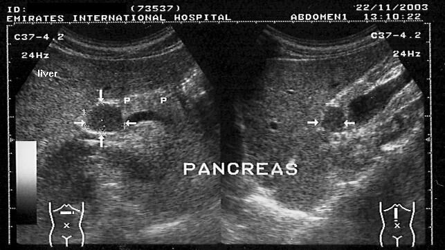
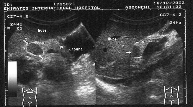
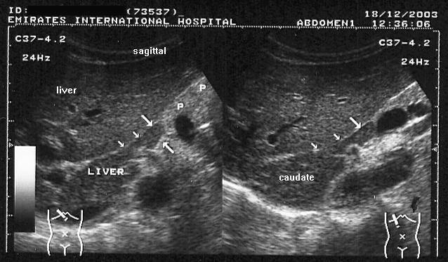
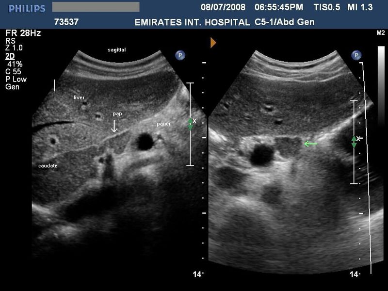
The papillary process of the caudate lobe of liver
1 2 3 4
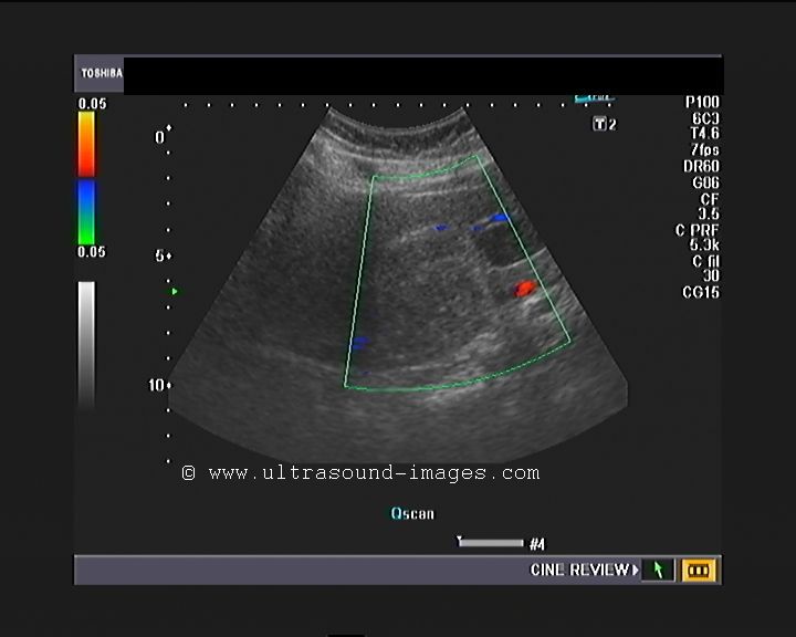
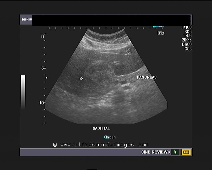
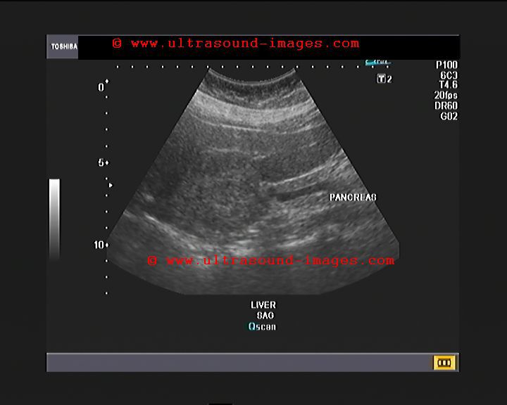
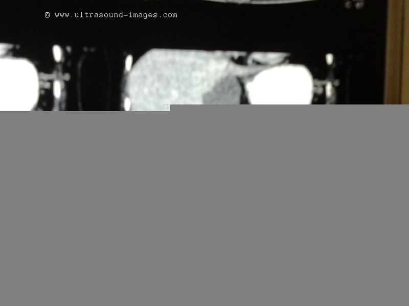
This patient presented with abdominal discomfort and underwent sonography of the abdomen. Ultrasound images show a rounded mass in the region of the pancreatic head and isthmus. It shows the same echogenicity as the liver(see 1 and 2). This suggested the possibility of a pancreatic mass, possibly malignant. However, images 3 and 4, reveal a different diagnosis- the possible "mass" appears to be an extension of the caudate lobe of the liver. These ultrasound images are diagnostic of "papillary process of the caudate lobe of liver."This normal variant may thus mimic pancreatic or preaortic lymph node masses.
Images courtesy of Dr. Ravi Kadasne, UAE.
Reference: http://radiology.rsnajnls.org/cgi/reprint/173/3/631.pdf (free article and images)-- rated excellent.
Riedel's lobe of liver
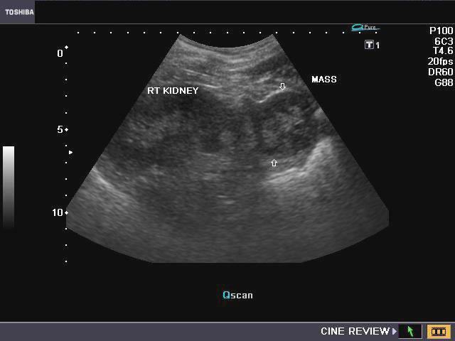
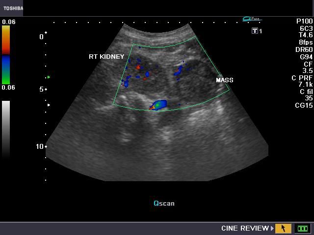
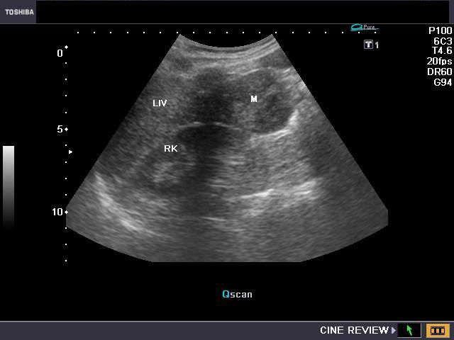
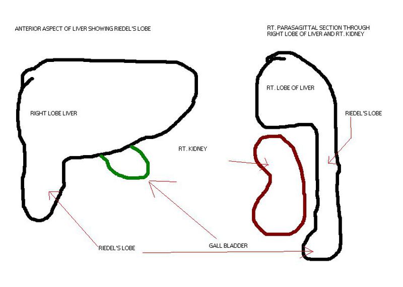
The above ultrasound images show what appears to be right renal lower pole mass (image in top row left). But further ultrasound imaging of the liver shows it to be an extension of the right lobe draped over the anterior surface of the right kidney. The apparent mass (M) appears to be on the tip of the tongue of liver tissue. This beak of liver tissue extending from the right lobe of liver is called Riedel's lobe of the liver and is a very rare normal variant. This patient was referred to a CT imaging center for further evaluation of the suspected mass in the tip of the Riedel's lobe. The sketch in bottom row-Right shows detailed anatomy of the Riedel's lobe. The gall bladder was visualized and appeared normal.
Reference: http://www.medcyclopaedia.com/library/topics/volume_iv_1/r/riedels_lobe.aspx
Elongated Left Lobe of Liver (the Beaver Tailed Liver)
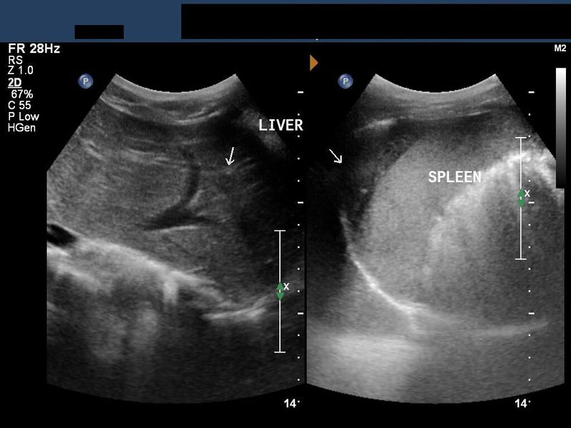

Long left lobe of liver (normal variant).
In certain thin individuals (usually seen in thin women), the left lobe of liver appears elongated (see ultrasound images above), and overlies the spleen. In the above pictures, the spleen is seen to be hyperechoic compared to the left lobe (the so called "beaver tailed liver"). Ultrasound images courtesy of Dr. Ravi Kadasne, UAE. The machine used here is the Philips IU 22.
Liver cyst
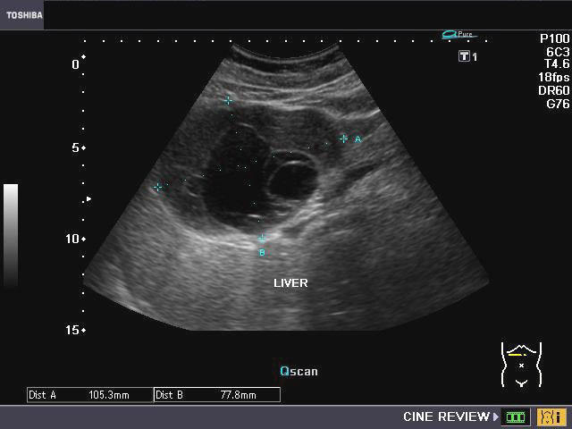
A) Sonography of hepatic cysts
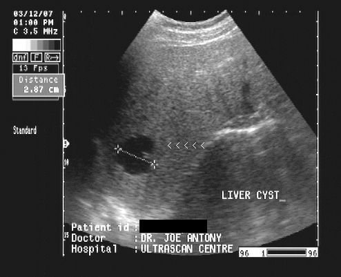
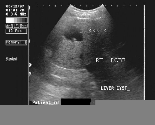
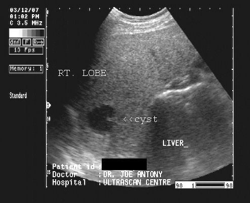
This elderly male patient came for routine sonography of the abdomen. Ultrasound images reveal an anechoic lesion in the right lobe of the liver, with thin walls and clear fluid content. A small projection is seen from the wall. This may be thickening of the wall or a fine nodule. These findings suggest a liver cyst. Differential diagnosis: biliary cystadenoma. Liver cysts are true cysts of the liver and have fluid content lined by epithelium. Images taken using a Pie Scanner 100 Falco.
Reference:
1)http://www.emedicine.com/med/topic2716.htm (free article and images)
2) http://www.emedicine.com/med/topic492.htm (free article)
B) Simple liver cyst: An unusual presentation
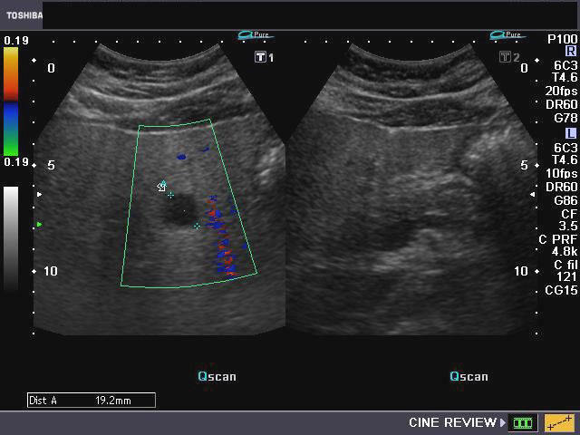


This middle aged male patient shows a cystic lesion of 1.9 cms. in the left lobe of liver on ultrasound imaging. However, what was interesting was the close proximity of the cystic lesion with a tributary of the portal vein. Color and Power Doppler images of the hepatic cyst showed no flow in the liver cyst. However, the spectral Doppler trace showed faint pulsations within this lesion. These pulsations appear to be transmitted from the abdominal aorta and appear insignificant. Thus we arrived at a diagnosis of a simple cyst of the left lobe of the liver, based on these ultrasound and Color Doppler images. Absence of flow in this cyst ruled out possibilities like a vascular lesion like aneurysm of the portal vein or AVM. The liver also shows moderate fatty change. This cyst was an incidental finding as the patient had no symptoms related to this lesion.
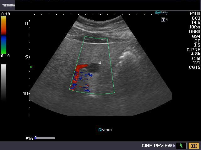
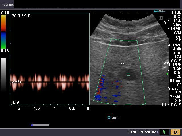
Hydatid cyst or echinococcosis of liver
This middle-aged female patient presented with non-specific abdominal pain. Ultrasound imaging of the liver shows a large cystic lesion with fine debris within it. There are also multiple cysts (daughter cysts) within the main cyst (parent cyst). Color and Power Doppler images of the cyst show few vessels along the rim of the parent cyst. These ultrasound images are diagnostic of Hydatid cyst (echinococcosis) of the liver. the fine debris in the cyst suggest hydatid "sand". WHO (World Health Organization) studies have classified the Cystic Echinococcosis or Hydatid Cyst into the following subtypes, based on their sonographic appearance: a) Cystic lesion- here there is a simple cyst in the affected organ. This appearance is not diagnostic for echinococcosis. b) Active cysts- here multiple cysts or septae are present in the parent cyst. c) Transitional stage-Daughter cysts may be present in the parent cyst with hydatid sand or debris within the cyst. (The case described here is in the transitional stage). d) The inactive stage- here the cyst is echogenic and may be partially or completely collapsed on itself.
Reference:
1)http://emedicine.medscape.com/article/216432-diagnosis
2) http://emedicine.medscape.com/article/178648-diagnosis
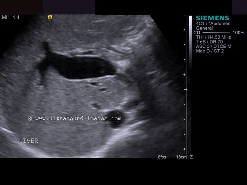
Liver metastases
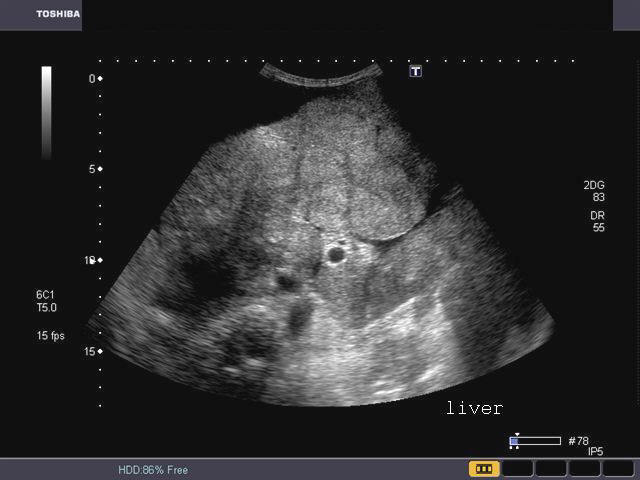
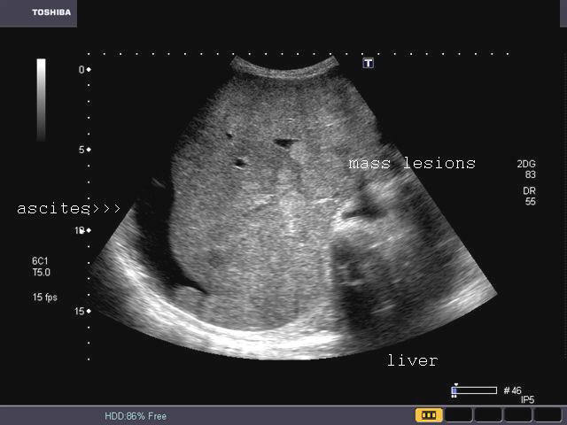
These ultrasound images of the liver reveal multiple, focal, echogenic mass lesions throughout the liver. The liver shows a nodular appearance. Such mass lesions may be due to hepatocellular carcinoma, Gastrointestinal malignancies or renal carcinoma. The more echogenic the masses, the masses are likely to be more vascular in nature. There is also associated pleural effusion and ascites.
Images taken using a Toshiba Xario machine. Images courtesy of Dr. Gunjan Puri, Surat, India.
Hepatic abscess or abscess of Liver (Amebic liver abscess)


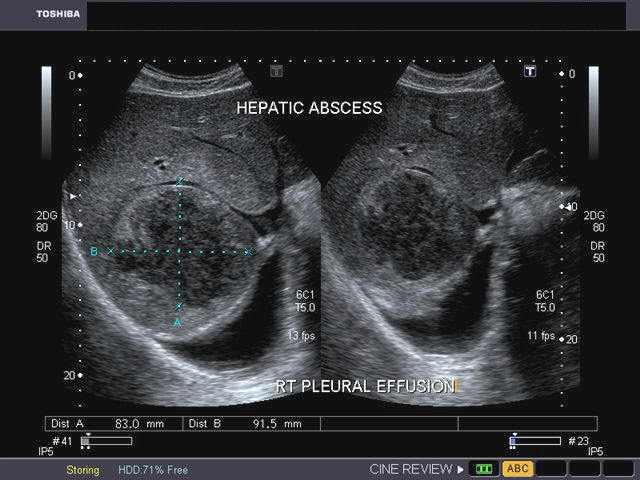
Sonography of the liver was done in this case of pain in the right iliac fossa. Ultrasound image on left shows thickened, edematous wall of the caecum s/o typhlitis. Image on right shows 2 hypoechoic lesions in the right lobe of liver. Both lesions are fairly large and show inhomogenous echotexture. In view of the underlying disease in the caecum, a diagnosis of typhlitis with amebic liver (hepatic) abscess (developing stage) was made.
In the above 2 ultrasound images of the liver, observe the markedly hypoechoic nature of the lesions suggesting further breakdown of the solid liver tissue ( liquifactive necrosis). On aspiration, the fluid was typically anchovy sauce like in appearance. This suggests hepatic amebic abscess at a more advanced stage. The gall bladder shows wall thickening suggesting edema of the wall. The image on right also shows a small pleural effusion just above the right lobe abscess (reactionary fluid collection due to pleuritis). (All 4 ultrasound images, taken using a Toshiba Xario ultrasound system, are courtesy of Gunjan Puri, MD, India).
Reference http://emedicine.medscape.com/article/188802-overview (free article).
Ruptured liver abscess (rupture or leak of hepatic abscess)
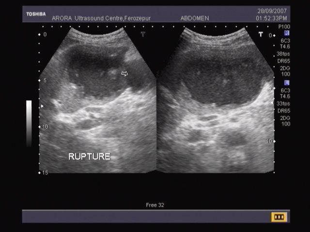
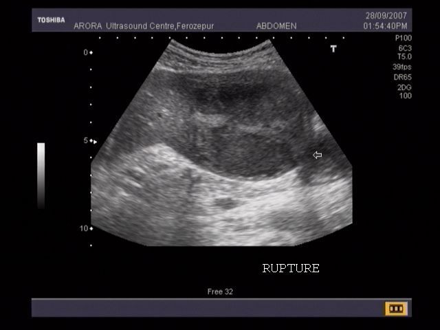

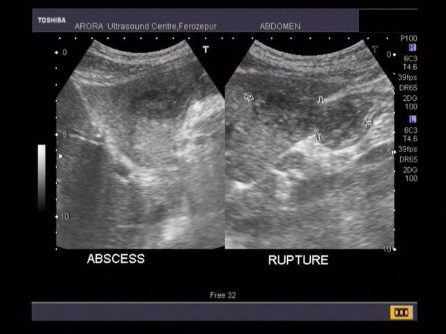

The above ultrasound images show a large abscess of the right lobe of liver with rupture of the lower margin of the abscess. The "leaked" fluid is contained within localized adhesions in the peritoneal cavity. The lowest image is a CT scan of the affected part. Amebic liver abscesses usually involve the right lobe of liver. Ultrasound images taken using a Nemio-30 Ultrasound system by Vikas Arora, MD, India.
Reference: Ultrasound imaging of hepatic abscess
Radiopedia- ruptured liver abscess
Hemangioma of liver (hepatic hemangioma)
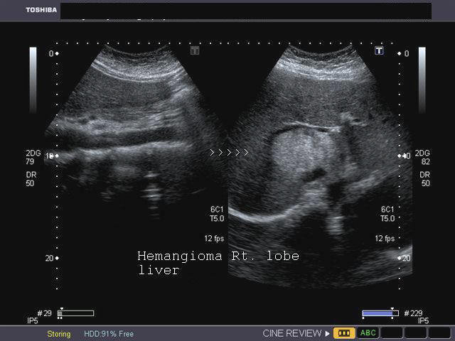
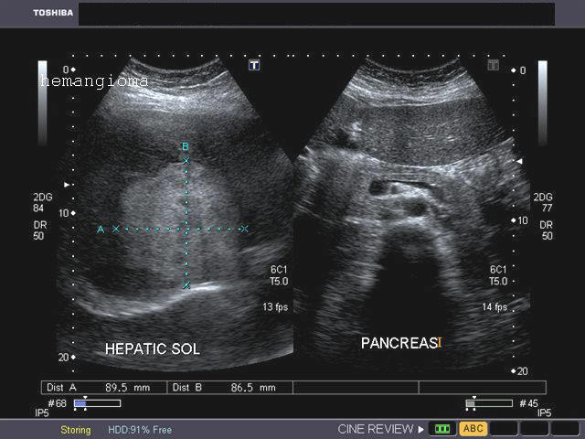
Sonography of the liver was done in this adult female patient. Images show a large (8 cms.) rounded, well defined, hyperechoic, non-calcific mass in the right lobe of liver. There is a moderate amount of acoustic enhancement posterior to the lesion. These ultrasound images show some of the typical features of hemangioma (cavernous) of the liver. In this case the patient is an adult female which is typical of hepatic hemangioma. However, usually hemangioma are smaller in size (less than 4 cms.). Ultrasound images were taken using a Toshiba Xario ultrasound system, courtesy of Gunjan Puri, MD, India.
Multiple Hemangioma of the Liver
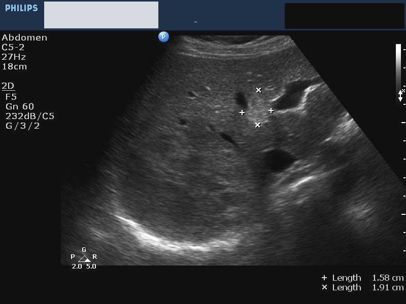
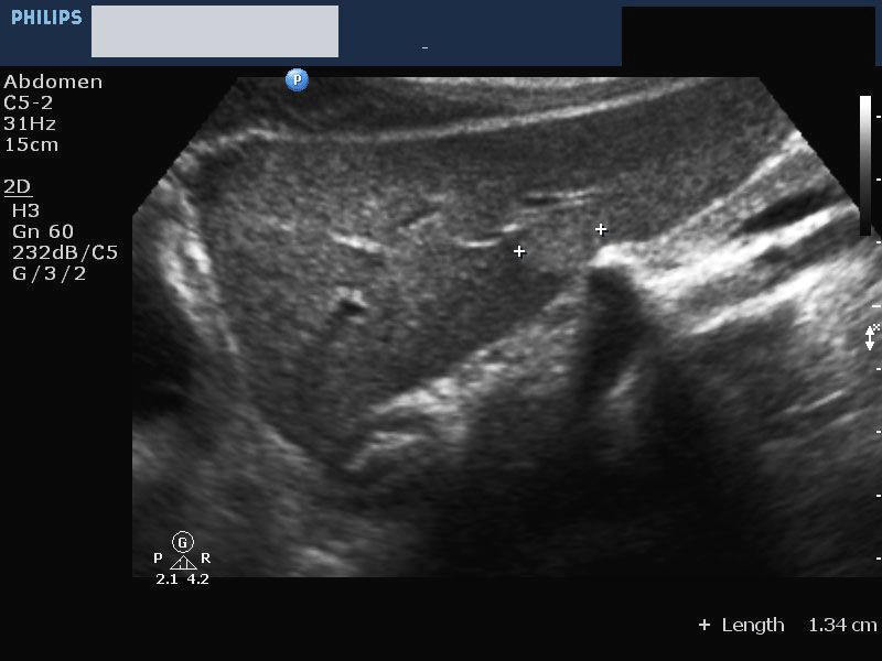
In the above 2 ultrasound images, there are multiple hemangioma of the liver. the lesions are small in size. Such lesions are rather atypical of hemangioma. These 2 ultrasound images are courtesy of Mr. Shlomo Gobi, Israel. The machine used here is the Philips HD 11 system.
Reference:http://emedicine.medscape.com/article/364860-overview (free article)
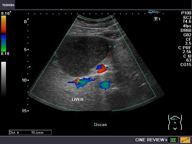
Hemangioma of the caudate lobe of liver
This middle aged lady has a large echogenic mass in the caudate lobe of liver (see ultrasound and color Doppler images above). Color Doppler images show poor flow despite the fact that this is a tumor of vascular origin.
CT images of the same mass


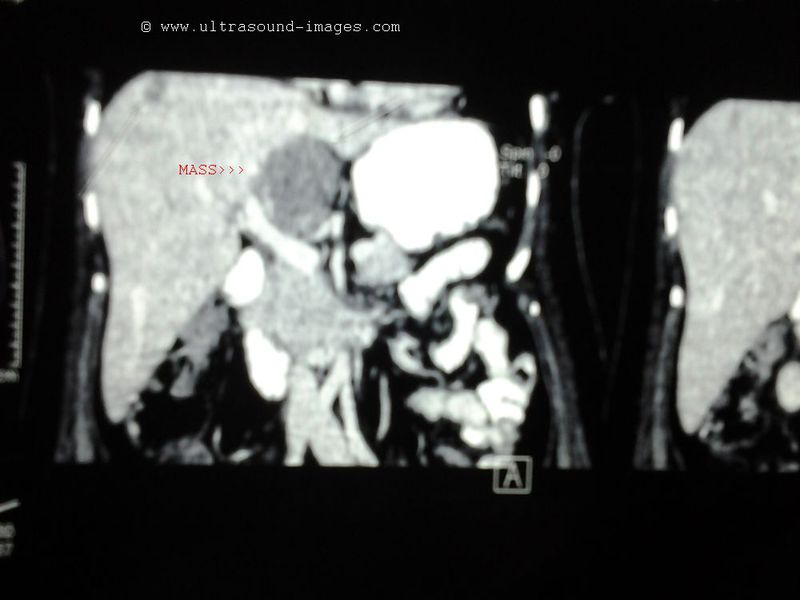
The CT scan images also confirm the presence of a large, hypodense mass in the caudate lobe which enhances (delayed) on post contrast study. These findings are typical appearances of hemangioma of the liver.
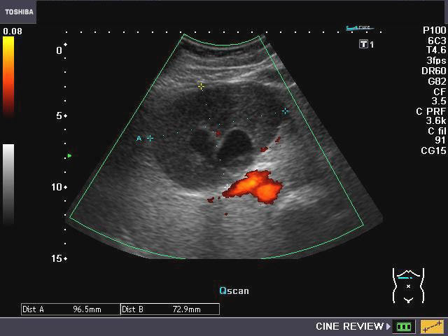
Portal vein stenosis in post liver transplant case
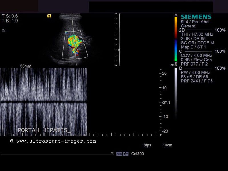
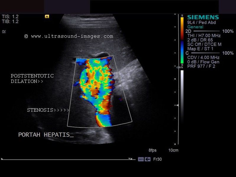


The above color Doppler and spectral doppler trace show a markedly turbrulent flow inside a dilated portal vein! But what is the reason for such turbulent and high velocity flows inside the portal vein? This patient is a child and is a case of live donor liver transplant.
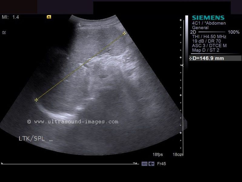
Observe the stenosis of the portal vein close to the porta hepatis. This is the site of anastomosis of the portal vein during the liver transplant procedure. The site of anastomosis of the portal vein between the donor and recipient is prone to undergo necrosis of the vein and consequent fibrosis with resultant stenosis. The poststentoic part of the portal vein shows marked dilatation and is due to the high velocity turbulent flow from the stenotic site. There is also evidence of splenomegaly in this child.
The above case and images are courtesy of Mr. Shlomo Gobi, Israel.
Reference: article on portal vein stenosis and its management
liver-calcification

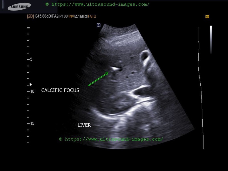
Liver calcification seen in right lobe of liver in the above ultrasound images:
= measures 0.7 cms. and may be due to calcified dead parasites or tuberculous infection
= calcification is also seen in certain liver tumors
= commonly such calcific foci in liver seen in parastic infestations as hydatids, schistosomes, histoplasmosis etc
References: calcification in liver- sonography
© 2019, Joe Antony, MD All images and content in this site are copyrighted. www.ultrasound-images.com When visiting this site, we may use cookies to improve the performance of this site. Use of this site means you accept the use of cookies. Read our privacy policy at this page: https://www.ultrasound-images.com/privacy-policy/